The need for new approaches for surgical treatment of medically refractory epilepsy is clear. Seizure-free outcomes for nonlesional, extratemporal epilepsy are only 40-50%. Our research suggests that using high-bandwidth EEG to map High Frequency Oscillations (HFOs) and Microseizures may better localize epileptic networks, and enable more effective surgery and antiepileptic device therapy. Our work in this area includes:
I. Using novel hardware to record high-bandwidth intracranial EEG (iEEG).
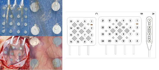
Example of high resolution iEEG grid to record from human neocortex. We use custom designed depth and subdural electrodes which are modified by the addition of micro-contacts to record high-resolution data that is of much higher spatial resolution than standard iEEG.
II. Unsupervised detection and classification of ictal patterns
We have validated machine learning methods for detecting and mapping epilepsy biomarkers in huge (Terabyte) data streams generated by high resolution arrays, and algorithms for detecting seizures with sufficient accuracy for an implanted human device.
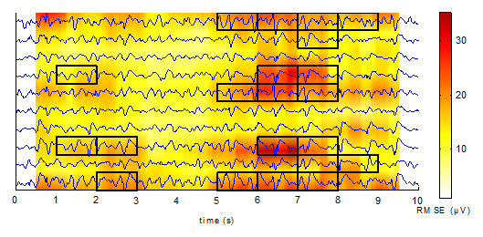
Anomaly visualizations of EEG using a novel algorithm. More anomalous areas of the signal are redder in color. Samples that a human reviewer independently labeled as non-background are boxed. J Neural Eng. 2011 Jun;8(3):036015.
III. Real-time intraoperative mapping of epilepsy biomarkers
At the Hospital of the University of Pennsylvania, high bandwidth iEEG data are collected while the patients are being monitored in the HUP Epilepsy Monitoring Unit (EMU) in parallel to our clinical monitoring equipment using a Neuralynx data acquisition and processing system. This is a sophisticated, FDA-approved system capable of collecting 256 channels of high-bandwidth multi-scale neurophysiologic data that enable us to map functional networks in the brain in great detail. The Neuralynx is coupled to a fast computer equipped with 32 graphical processing units (GPUs) to perform detection, localization and mapping of epilepsy biomarkers on 3-D brain image reconstructions morphed to intraoperative photographs (Khambhati, et. al. in review).
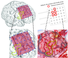
Figure demonstrating electrode grid and HFO map morphed to 3-D brain reconstruction from intra-operative photograph. (Khambhati et al., in review)
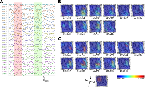
Figure demonstrating iEEG segment during the interictal period (A), 3-D map demonstrating HFO spatial map during the interictal (between seizure) period (B), and as a seizure approaches (C). Khambhati et al., in review.